ImmunoFluor™-Maleimide (PEGylated) (Post-insertion)
Description
During the past five decades, various types of chemistries have been used for conjugation of molecules such as antibodies to the surface of the liposomes. In general, the conjugation can be achieved through the N-terminus, the C-terminus or the available sulfur (e.g. Fab’ fraction or thiolated Ab). Not all chemistries have the same yield and efficiency of conjugation and often reproducing biocompatible batches can be a challenge. Coupling of sulfhydryl groups with maleimide groups has been the most widely used conjugation of antibodies to liposomes. Different lipids which are offered for thioether conjugation contain maleimide, aromatic maleimides such as N-[4-(p-maleimidophenyl)-butyryl] (MPB) or 4-(N-maleimidomethyl)cyclohexane-1-carboxylate (MCC) group. The maleimide function group of MCC which contains an aliphatic cyclohexane ring is more stable toward hydrolysis in aqueous reaction environments rather than the aromatic phenyl group of MPB. MPB and MCC lipids are non-PEGylated lipids and they have separate kits and protocols than PEGylated maleimide lipids.
One of the major problems of using maleimide chemistry for conjugation is the rapid hydrolysis of maleimide lipid. The rate of hydrolysis is much faster in alkaline pH and therefore controlling the pH throughout the entire process is necessary and it is recommended to use the pH of 7. Due to the hydrolysis of maleimide group, our kits are designed for post-insertion of ligand conjugated maleimide lipid into the preformed liposomes. After post conjugation the liposomes have to be used right away because hydrolysis may occur after sulfhydryl coupling to the maleimide as well. Another problem is the reactivity and oxygen sensitivity of sulfhydryl group on thiolated antibody or Fab’ fragment. Due to that the conjugation reaction should be done under argon or nitrogen using inflatable polyethylene glovebag chambers.
Thiolation which is adapted to the modification of all of the antibody functional groups, is relatively clean, fast, and efficient. However, different antibodies may be more sensitive to some procedures than others. Therefore, it is recommended to select the chemistry and site of modification depending on what procedures are compatible with the antibody.
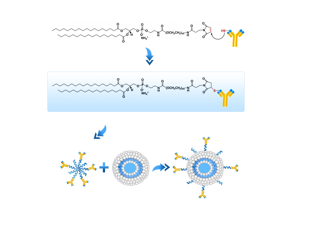
ImmunoFluor™-Maleimide is a PEGylated product. For other sulfhydryl reactive (PEGylated and non-PEGyalated products) and also ImmunoFluor™ products suitable for other types conjugation methods see here.
Formulation Information
ImmunoFluor™-Maleimide (PEGylated) (Post-insertion)
| Post-insertion Kit (3 Vials) | Specification |
|---|---|
| Vial 1 | Preformed liposomes composed of HSPC:Cholesterol:Fluorescent lipid (59.5:40:0.5 molar ratio) |
| Vial 2 | DSPE-PEG(2000)-Maleimide lipid (reactive PEGylated lipid) in powder form |
| Vial 3 | DSPE-PEG(2000) lipid (non-reactive PEGylated lipid) in powder form |
| Lipid Composition for Vial 1* | Concentration (mg/ml) | Concentration (mM) | Molar Ratio Percentage |
|---|---|---|---|
| Hydrogenated Soy PC | ~11.5 | ~14.66 | ~60 |
| Cholesterol | 3.83 | 9.9 | 40 |
| Fluorescent Lipid (see below) | Varies based on the dye | Varies based on the dye | Varies based on the dye |
| Total | ~15.33 mg/ml | ~24.56 mM | 100 |
| * For the 5-ml kit, the volume of vial 1 is 4 ml. 1 ml of micelle solution that are formed using vials 2 and 3 will be added to this vial to make the final volume of 5 ml in the final product. For the 2-ml kit, the volume of vial 1 is 1.6 ml. 0.4 ml of micelle solution that is formed using vials 2 and 3 will be added to this vial to make the final volume of 2 ml in the final product. | |||
| Fluorescent Dye | Excitation/Emission (nm) | Molecular Structure |
|---|---|---|
| 1,1'-Dioctadecyl-3,3,3',3'-tetramethylindocarbocyanine perchlorate (DiI) | 549/565 | 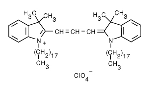 |
| 3,3'-Dilinoleyloxacarbocyanine perchlorate (DiO) | 484/501 |  |
| 1,1'-Dioctadecyl-3,3,3',3'-tetramethylindodicarbocyanine, 4-chlorobenzenesulfonate salt (DiD) | 644/665 | 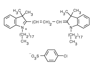 |
| 1,1'-Dioctadecyl-3,3,3',3'-tetramethylindotricarbocyanine iodide (DiR) | 750/780 | 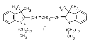 |
| 4-(4-(Dihexadecylamino)styryl)-N-methylpyridinium iodide (DiA) | 456/590 |  |
| 1,2-Distearoyl-sn-glycero-3-phosphoethanolamine-N-(7-nitro-2-1,3-benzoxadiazol-4-yl) (ammonium salt) (NBD on head group) | 460/535 |  |
| 1-Palmitoyl-2-{12-[(7-nitro-2-1,3-benzoxadiazol-4-yl)amino]dodecanoyl}-sn-glycero-3-phosphocholine (NBD on fatty acid tail) | 460/534 |  |
| 1,2-Dipalmitoyl-sn-glycero-3-phosphoethanolamine-N-(lissamine rhodamine B sulfonyl) (ammonium salt) (Rhodamine lipid) | 560/583 | 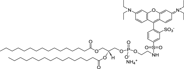 |
| 1,2-Dioleoyl-sn-glycero-3-phosphoethanolamine-N-(5-dimethylamino-1-naphthalenesulfonyl) (ammonium salt) (Dansyl lipid) | 336/513 |  |
| 1,2-Dipalmitoyl-sn-glycero-3-phosphoethanolamine-N-(1-pyrenesulfonyl) (ammonium salt) (Pyrene lipid) | 351/379 |  |
| Buffer and Liposome Size for Vial 1 | Specification |
|---|---|
| Buffer | Phosphate Buffered Saline |
| pH | 7.4 |
| Liposome Size | 100 nm |
| Vial 2 * | Specification |
|---|---|
| DSPE-PEG(2000)-Maleimide Lipid | This vial contains reactive DSPE-PEG(2000)-Maleimide lipid in powder form. This lipid is conjugated to a reactive protein, peptide or ligand containing sulfhydryl and then mixed with non-reactive DSPE-PEG(2000) lipid in aqueous solution to form micelles. The PEGylated lipid micelles are incubated with preformed liposomes in vial 1 and PEG lipids will post-insert themselves into the liposomes.  |
| * The amount of the powdered PEG(2000)-Maleimide lipid for 2-ml kit is 1.34 mg and for 5-ml kit is 3.34 mg. | |
| Vial 3 * | Specification |
|---|---|
| DSPE-PEG(2000) Lipid | This vial contains non-reactive DSPE-PEG(2000) lipid in powder form. This lipid in mixed with DSPE-PEG(2000)-NHS lipid which is already conjugated to a ligand (protein, peptide, etc.) in aqueous solution to form micelles. The PEGylated lipid micelles are incubated with preformed liposomes in vial 1 and PEG lipids will post-insert themselves into the liposomes. |
| * The amount of the powdered PEG(2000)-DSPE lipid for the 2-ml kit is 5 mg and for the 5-ml kit is 12.5 mg. | |
Conjugation Protocol (Post-insertion)
Materials and Equipment
The 3-vial post-insertion kit contains preformed liposomes (vial 1), DSPE-PEG(2000)-Maleimide lipid in powder form (vial 2) and non-reactive PEGylated lipid in powder form (vial 3). In order to use the post-insertion kit, you will need:
- Two small 10-ml round bottom flasks or two small glass vials
- A rotary evaporator. We understand that many labs might not have a rotovap. Alternatively, you can use a nitrogen tank connected to a thin hose for creating a stream of nitrogen flow to dry the lipid and make a thin film.
- A small amount of a solvent such a chloroform or methylene chloride (you will only need a few milliliters).
- Phosphate buffered saline (PBS). pH should be adjusted to 7.
- 2-Mercaptoethanol.
- Aldrich®-Atmosbag connected to a nitrogen tank. Due to oxygen sensitivity of the reaction, the coupling reacting should be done in oxygen-free environment.
- Float-A-Lyzer® with a proper MWCO that easily allows the cleanup of your liposome conjugated ligand from free and non-conjugated protein/peptide/ligand. You need to make sure that the MWCO is below 1,000,000 dalton. At 1,000,000 dalton, the pore size on the dialysis membrane gets close to 100 nm and therefore your liposomes can be dialyzed out. You cannot use dialysis cassettes blindly. Please understand the technique before using either spin columns or dialysis cassettes. If you do not use the correct MWCO, you can lose your entire prep. For this protocol, we recommend MWCO of 300,000 dalton.
- A Sonicator. It is better to have a bath sonicator. It you do not, that is fine. You still can follow the protocol. You may also use a vortex instead of the sonicator for agitation of the solution as well.
Preparation Method
- Dissolve the content of vial 3 (non-reactive PEGylated lipid) in 100 µl of chloroform or methylene chloride. Transfer the solution to a 10 ml round bottom flask. Dry the chloroform using a rotary evaporator or under a stream of nitrogen in order to make a dried lipid film.
- Add 100 µl of PBS buffer to the dried lipid film. It is preferred to sonicate the hydrated lipid film using a bath sonicator and sonicate the micelle solution for 5 minutes. If you do not have a bath sonicator then hydrate the dried lipid film with PBS for at least 1 hour and constantly rotate the solution in the round bottom flask using a rotavap (not connected to vacuum) or by hand to make sure that all the dried lipid on the wall of the round bottom flask will go to the solution and form micelles. Alternatively, you can use a vortex to agitate the solution. The goal is to have all the dried lipid on the wall of the round bottom glass to go to the micelle solution. Cover the mouth of the round bottom flask with parafilm. Refrigerate the micelle solution of non-reactive PEG lipids until it is ready to be mixed with micelles formed in the step 5.
- The kit contains 1.30 mg (0.22 µmol) of reactive DSPE-PEG(2000)-Maleimide lipid (vial 2). Transfer the solution to a 10 ml round bottom flask. Dry the chloroform using a rotary evaporator or under a stream of nitrogen to make a dried lipid film.
- Dried DSPE-PEG-Maleimide film is hydrated with PBS buffer to form a micellar lipid solution. Hydrate the 1.30 mg of dried DSPE-PEG-Maleimide lipid film in 100 µl of buffer.
- Incubate the micellar lipid solution with the antibody, protein or peptide at 3:1 molar ratio or lipid to protein. Allow the reaction to proceed in phosphate buffer under the nitrogen (inert gas) chamber for 8 hours at room temperature with moderate stirring. The concentration of antibody, peptide or protein that is added to micellar solution is depend on the solubility of your molecule. It is recommended to use a fairly concentrated solution. A volume around 100 µl of antibody, peptide or protein is used for our 2-ml kit.
- The excess maleimide groups were capped by reaction with 2-mercaptoethanol. The reaction is quenched with 2 mM 2-mercaptoethanol for 30 min.
- The micelles obtained from the steps 2 and 5 are mixed. Total volume of the 2 mixed micelles is 300 µl. Incubate the mixed micelles with preformed liposomes (vial 1) at 60℃ for 30 min.
- Remove non-conjugated antibody, protein, peptide or ligand by dialysis. We prefer dialysis to size exclusion columns. Dialysis is a much slower process but there will be minimum loss of immunoliposomes after the prep is cleaned from non-conjugated protein/peptide/ligand. Spin columns are much faster, but you can easily lose over 50% of the liposomes on the spin column. We recommend using Float-A-Lyzer® dialysis cassette from Spectrum Labs. You need to choose a cassette with proper MWCO depending on the MW of your protein, ligand, antibody or antibody fragment. In this case we recommend using a dialysis cassette with MWCO of 300,000 dalton. NOTE: If you decide to use a dialysis cassette you need to make sure that the MWCO is below 1,000,000 dalton. At 1,000,000 dalton, the pore size on the dialysis membrane gets close to 100 nm and therefore your liposomes can be dialyzed out. You cannot use dialysis cassettes and spin columns blindly. They come in various sizes and you need to choose the correct size wisely. Dialyze the immunoliposome solution in 1 liter of PBS at pH 7 for 8 hours. Change the dialysis buffer with a fresh 1 liter of PBS and let is dialyze for another 8 hours. After this step, your cleaned up immunoliposome is ready to be used.
Quantification of reactive sulfhydryl in antibodies or ligands (Ellman’s Assay)
The yield of conjugation is the most important factor in formulating immunoliposomes. Many scientists simply assume that their thiolated antibody or the Fab’ fraction contains reactive sulfhydryl for conjugation to maleimide lipid without further assaying. Disulfide bridge can form very easily so it is very important to quantify the available reactive sulfhydryl in your antibody or ligand solution before performing the conjugation reaction with maleimide liposomes.
Ellman’s assay is a widely used assay for determining the amount of free sulfhydryl. You can follow the step by step protocol here.
Liposome Particle Calculator
ImmunoFluor™ liposomes are unilamellar and sized to 100 nm. The molar concentration of liposome is 24.56 mM. By having liposome diameter (nm) and lipid concentration (µM), you can calculate the total number of the lipids in one liposome and the number of the liposomes in one milliliter of the liposome solution. To use the calculator click here.
Technical Notes
- After conjugation reactions, liposomes containing excess maleimide or thiol groups may exhibit undesirable qualities, such as aggregation, reactions in vitro and in vivo, and immunogenicity. These reactive moieties can be quenched with reagents containing iodo-, maleimide, or sulfhydryl groups where appropriate. This is likely to be a particularly serious problem for thiolated liposomes. Therefore, it is recommended that the antibody be thiolated in order to generate the appropriate reactive entities for the final conjugation reaction.
- In order to prevent oxidation of sulfhydryl on antibody and formation of disulfide bridge, the coupling reaction must be performed under an inert atmosphere such as argon or nitrogen. To set up a inert gas chamber we recommend using Aldrich®-Atmosbag with is a flexible, inflatable polyethylene chamber with built-in gloves which is a portable and inexpensive alternative to laboratory glove box.
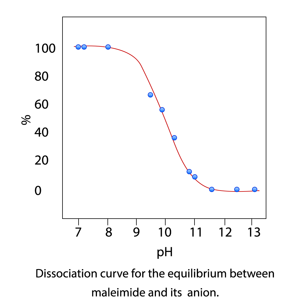
- Maleimide group on lipid is highly sensitive of alkaline pH and it will hydrolyze rapidly at higher pH. Experimental investigations have been shown that in alkaline condition (pH > 7.5), maleimide and its derivatives are hydrolyzed to a non-reactive maleamic acid (see the figure below). This instability should be taken into account in any quantitative procedures, such as coupling with sulfhydryl groups. Therefore, it is very important to make sure that the pH of the reaction with stay between 6.5 and 7 during the entire process.
Database
Direct link to the database page for easy navigation: Immunoliposomes Conjugation Database
Appearance
ImmunoFluor™-Maleimide (PEGylated) post-insertion kit comes in three vials: vial 1 formulation is colored and the color depends on the type of the fluorescent dye that is used (see SDS for appearance). It contains nano size unilamellar liposomes which does not contain any reactive of non-reactive PEGylated lipid. Usually due to the small size of liposomes no settling will occur in the bottom of the vial. Vial 2 contains reactive DSPE-PEG(2000)-Maleimide lipid in white powder form. Vial 3 contains non-reactive DSPE-PEG(2000) lipid in white powder form.
Ordering/Shipping Information
- All liposome based formulations are shipped on blue ice at 4 C in insulated packages using overnight shipping or international express shipping.
- Liposomes should NEVER be frozen. Ice crystals that form in the lipid membrane can rupture the membrane, change the size of the liposomes and cause the encapsulated drug to leak out. Liposomes in liquid form should always be kept in the refrigerator.
- Clients who order from outside of the United States of America are responsible for their government import taxes and customs paperwork. Encapsula NanoSciences is NOT responsible for importation fees to countries outside of the United States of America.
- We strongly encourage the clients in Japan, Korea, Taiwan and China to order via a distributor. Tough customs clearance regulations in these countries will cause delay in custom clearance of these perishable formulations if ordered directly through us. Distributors can easily clear the packages from customs. To see the list of the distributors click here.
- Clients ordering from universities and research institutes in Australia should keep in mind that the liposome formulations are made from synthetic material and the formulations do not require a “permit to import quarantine material”. Liposomes are NOT biological products.
- If you would like your institute’s FedEx or DHL account to be charged for shipping, then please provide the account number at the time of ordering.
- Encapsula NanoSciences has no control over delays due to inclement weather or customs clearance delays. You will receive a FedEx or DHL tracking number once your order is confirmed. Contact FedEx or DHL in advance and make sure that the paperwork for customs is done on time. All subsequent shipping inquiries should be directed to Federal Express or DHL.
Storage and Shelf Life
Storage
ImmunoFluor™ products should always be stored at in the dark at 4°C, except when brought to room temperature for brief periods prior to animal dosing. DO NOT FREEZE. If the suspension is frozen, the encapsulated drug can be released from the liposomes thus limiting its effectiveness. In addition, the size of the liposomes will also change upon freezing and thawing.
Shelf Life
ImmunoFluor™-Maleimide kit is made on daily basis. The batch that is shipped is manufactured on the same day. It is advised to use the products within 2 months of the manufacturing date.


